Saturday, 28 November 2015

Field Trip: Aquascape Paradise

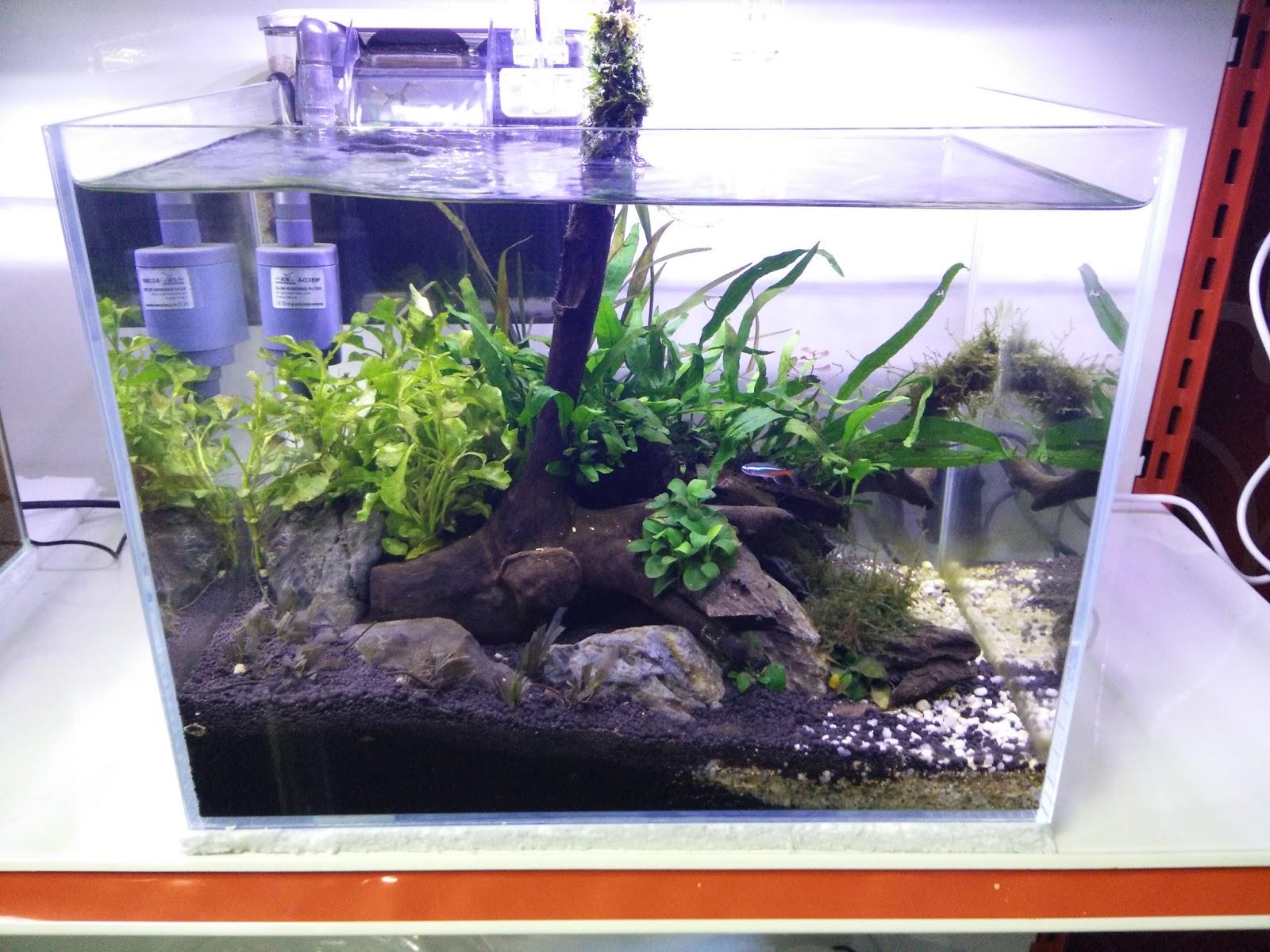
Aquascape is the arts of arranging aquatic plants as well as rock, stones, driftwood and so on. Other people call it as underwater gardening. Nomally, an aquarium would be decorate with fish plants and all sorts of creative idea that can be come out with. With our field trip to Aquascape Paradise we learned various ways to make underwater art using freshwater macrophytes that are truly outstanding.

Commonly used species are Blyxa sp., Myriophyllum sp., Hydrilla verticillata, Alternanthera sp., Anubias nana, Bacopa carolinianna, Hydrocotyle verticillata, H. leucocephala, Hemianthus sp.,  Java fern, Java moss and others which includes about 114 local species and imported species to create underwater concept. 

Materials that can be used
Driftwood

Rock imported from japan which have extra nutrient

Designs of the aquascape
There are different design outside there mostly based on japan and dutch


Paludariums: 
An aquarium that combines water and land inside the same environment.
Jungle style:
Natural, untrimmed look that provide wild appearance

Iwagumi style:
Main layout materials are stones and covered with mosses


Dutch style
By having multiple plants with different colour, and the tank have more than 80% covering the aquarium floors

Nature style:
Mountain like landscape

Types of aquatic plants
Mosses tied on the driftwood that resembles a tree


Terrarium

Example of how the shop grow their aquatic plants


Water are sprinkled from above to keep the soil and plant moisture


Special thanks to the management of Aquatic Paradise for the warm welcome and knowledge on aquascaping.

Practical 5: Propagation of Seaweed in Culture Media

Introduction
Seaweed are know to reproduce through fragmentation where if some parts of its thallus are torn or broken apart, they are able to regenerate and grow a new thallus. This is process is also carried out symbiotic with bacteria that supplies nutrients such as nitrogen to the seaweed. This study is to observe the species of bacteria that assist in fragmentation.

Objective
To determine which bacteria is best for the culturing the seaweed

To learn ways to propagate the seaweed

Material and Methods
Picture 1:Cut 1 cm of the seaweed


Picture 2: Rinsed the seaweed by using filtered sea water and shaken vigorously to sterilized the seaweed

Picture 3: Record the wet weight of seaweed


Picture 4: Inserted the culture media Von Stosch's Enrichment (VSE) and Provasoli Enrichment Seawater (PES)


Picture 5: Inserted the bacteria ( 4 types of bacteria)

Picture 6:Insertion of bacteria to algae sample.

Picture 7: Sealed the side of the container using parafilm

After that, incubate the seaweed for a photoperiod of 12 hr light and 12 hr dark with aeration


Results
Picture 8: First day of propagation

Picture 9: One week after the propagation
Picture 10: Few week after the propagation the buds begin to emerge
Discussion
The first week does not shows much growth or we can say probably no growth as there no new shoots growing out, however, the result can be seen after a few weeks as the shoot grow as long as 1 cm as shown in BAC3 which shows positive budding as the algae is associated with BAC3.

Precious Underwater Forest in Merambong Shoal, Johor


Merambong Shoal. What do we know about it? It is located at Gelang Patah, Johor. This mangrove island used to be a mainland for seagrasses all the way from Tanjung Adang Laut until Merambong. Seagrass species such as Halophila ovalis, Halophila minor, Halophila sp., and Halodule pinifolia are the main diet of our endangered animal, Dugong. The beautiful green underwater view has gone disappeared thanks to some thoughtless coastal development in 2002.

Image source from The Star

The land development not only slaughtered many seagrass species there, they also caused home for many other animals besides dugong such as turtles and seahorses. The enraging of a few of NGOs never seems bother the developer to stop the development.

However, the dark cloud has came to found a silver lining when some researcher found a recovery of a few species there where they saw the growth of the seagrasses there,

People must be aware of  the importance of having a seagrass bed to fisheries and also towards the ecosystems in order to build a greater future that applies eco-friendly activities.


Monday, 2 November 2015

Practical 1: Microalgae Culture Medium

Introduction

Microalgae or microphytes are actually the algae but in micro size that do not have roots or leaves. They can live in either freshwater or seawater depending on the species. They are unicellular species. As a species of algae, they are the primary producers that supply oxygen and food. Beside that, microalgae are a source of a wide variety of nutrients such as omega-3 fatty acids, and are a particularly large source of protein. In the wild,  they are eaten by mollusc, crustaceans and fish fry. Microalgae source of food are from minerals such as nitrates and phosphate. For example, some microalgae are Spirulina, chlorella, and blue-green algae, given to us (human) as nutrient supplement. Currently, there are findings prove that microalgae can be used to solve pollution problems, cleaning waste water while producing important goods for society. Therefore, preparing a suitable medium will allow us to better understand our selected microalgae species.


Objective
To learn how to use different equipment to sample the microalgae

To learn the optimum abiotic factors for microalgae to survive

To be able to prepare suitable algae agar medium

Material and Method

Different microalgal sampling equipments :

Basic Apparatus:
  • ·         250ml conical flask
  • ·         500ml Beaker
  • ·         Mixer
  • ·         pH meter
  • ·         Magnetic stirrer

Materials:

Bold's Basal Medium (BB)


f/2 Medium


f/2 + Si (Guillard's medium for diatoms)


Provasoli Enriched Seawater Media (PES)


Von Stoch Enrichment (VSE)


Macrophyte Medium Culture : Murashige and Skong (MS) Media 


Discussion
Each culture medium is specially formulated to best suit a group of microalgae for instance, f/2 is made for various microalgae while f/2+silicate is made for Bacillariophyta as they require silica to incorporate their cilica cell wall. While Bold Basal's Medium is one of the commonly used medium for freshwater macrophytes. Other mediums such as PES, VSE and MS are formulated for saltwater microalgae.

Conclusion
Various medium is used to best satisfy the growth of both freshwater and saltwater flora. The selection of a culture medium greatly depends on the type of nutrient required for a specific species of microalgae. The various agar mediums are also able to inhibit the growth of unwanted microalgae such as f/2 has no silica which does not promote the growth of diatoms. Therefore, it is best to study the nutrient requirement of each microalgae species when electing an agar medium.

Practical 2: Microalgae Identification


Introduction
Micro algae exist in a diverse marine ecosystem along with others of its species which includes those from Class Pheophyta, Rhodophyta and Chlorophyta in the same area along with other classes such as diatoms and. When starting a single species culture, it is essential to properly isolate a single species to produce a high quality culture that is not effected by competition with other species. This can be done through various techniques such as serial dilution, streak plating and also single cell micro pipetting.

Objective
To identify and classify the different types of microalgae found


To be able to use various methods of algae isolation which includes micropippette, streaking and dilution technique.

Material
Wire loops, Bunsen burner, Petri dish, 10x test tube, test tube rack, sterile screw-capped, Media, Agar

Method
Serial dilution
1.       Insert 9ml of media into each of ten test tubes with sterile 10ml pipettes.
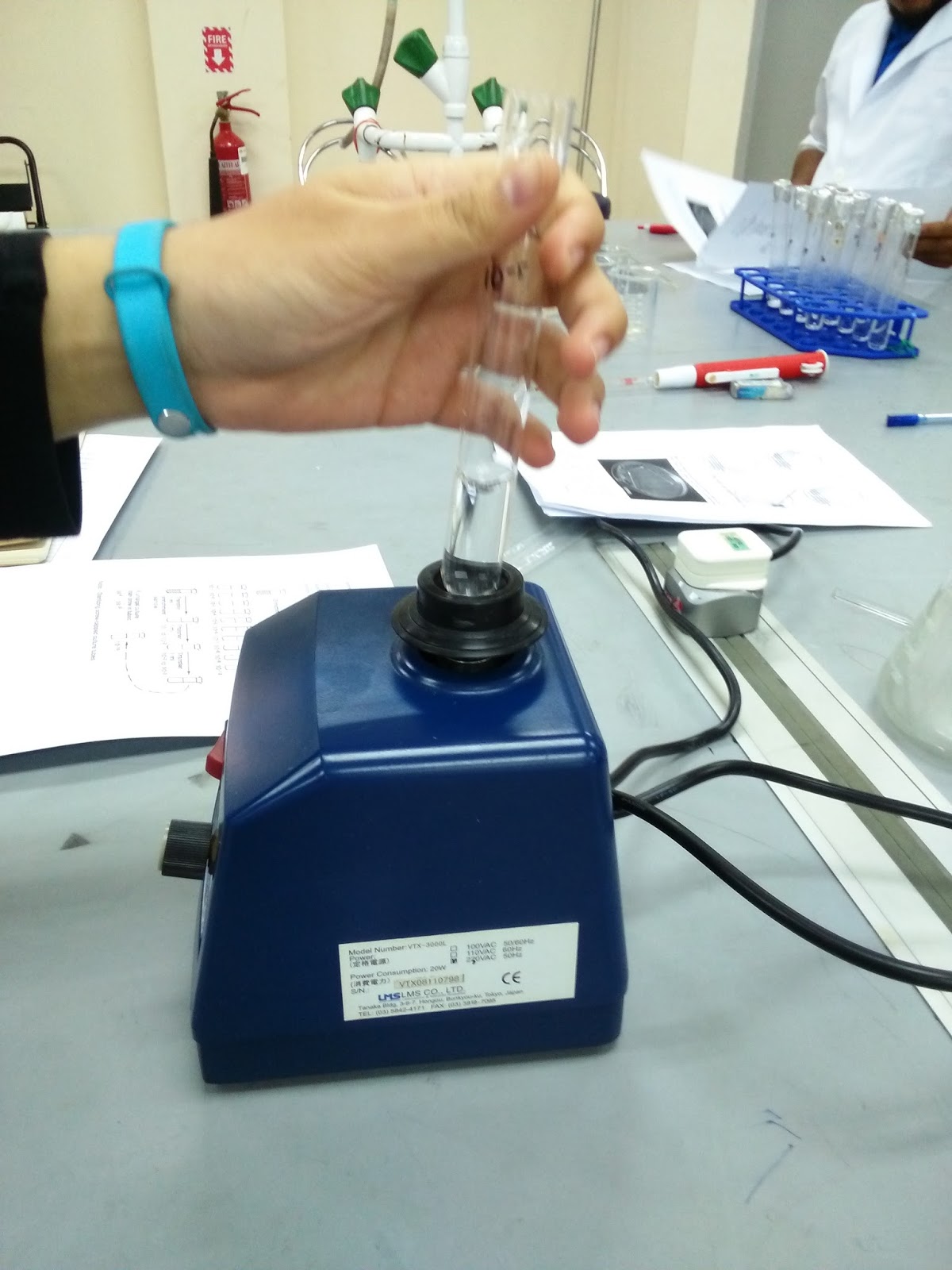
2.       Label the test tube from 10-1 until 10-10 to show the different dilution.
3.       Then, add 1ml of  enrichment sample to the first test tube which is (10-1) and shake gently.
4.       Take the 1ml of (10-1) dilution and add to the next test tube (10-2) and shake gently.
5.       Repeat the procedure by inserting to the other test tube (10-3 to 10-10).
6.       Incubation test tube is needed for controlled temperature and light conditions (put under florescent light)

Streak plating
1.       Prepare petri dishes that contain growth medium solidified with 1.5% of agar medium.
2.       The agar should not be more than 2/3 of the petri dish.
3.       Place 2 drops of phytoplankton sample at a spot at the edge of the petri dish.
4.       Sterilize the wire loop by using flame.
5.       By using aseptic technique, use the sterile wire loop, drag the drops of phytoplankton sample and make parallel streak ( Each streak have 4 line, in total 16 are needed)
6.       The force must not be too powerful which using the sterile wire loop as the phytoplankton sample are meant to be distribute on the surface of the agar.
7.       Invert and incubate under low light at constant temperature.


8.       Next week, the phytoplankton sample are being choose and observe under microscope to identify if the pure culture are successful and to detect invasive bacteria or phytoplankton.

Single cell micropipette
-A drop of culture X was placed onto a glass slide and observed under a compound microscope.
-A single species microalgae was selected and micropipetting is done to seperate the microalgae from the rest.
-Once a single microalgae is separated, it was placed onto a tray along with a culture medium to grow.

Results

        Image 1: Brown spots showed the presence of Amphora sp. in f/2+silica A agar plate.
Image 2: Brown colonies of Amphora sp and small green colonies of Tetraselmis sp in agar f/2+silica B.


Image 3: Only a single colony of Amphora sp in agar f/2 A.
Image 4: Green colonies of Tetraselmis sp and very few brown colonies which is Amphora sp in agar f/2 B

Image 5: Amphora sp observed under compound microscope.


Discussion
It is observed that streaking method was a success with multiple colonies growing. When observed under a compound microscope, Amphora sp can be found in both f/2 and f/2+silica agar plate though it requires silica to grow. However, Tetraselmis sp was also found in some of the medium, most are found in f/2 agar plate. The serial dilution gave us no results after a week as there might be a small amount of inoculum to begin with and their growth is very slow. Other than that, micropipetting method was successful in selecting only one species of microalgae; however, the medium dried out and the selected microalgae which was Tetraselmis sp had died off.
Conclusion
Various methods can be done to easily separate a single microalgae species to be cultured. The most effective method is micropipetting as we are able to select the species that we want but it takes time and requires skills to be done correctly.


Reference
http://www.marine.csiro.au/microalgae/methods/microalgal%20isolation%20techniques.htm

Appendix
image 6: Micropipetting

Image7: Carefully sucking selected microalgae.

Image 8: A mix microaglae culture known as Culture X

Image 9: Serial dilution technique.

Image 10: Mixing the microalgae and medium from serial dilution.


Image 11: Centrifuge used to produce pallets of Culture X
Image 12: Sterilising the wire loop before inoculating the pallet onto the agar plate.

Image 13: an f/2 medium agar plate after agar streaking of Culture X.