Introduction
Micro algae exist in a diverse marine ecosystem along with others of its species which includes those from Class Pheophyta, Rhodophyta and Chlorophyta in the same area along with other classes such as diatoms and. When starting a single species culture, it is essential to properly isolate a single species to produce a high quality culture that is not effected by competition with other species. This can be done through various techniques such as serial dilution, streak plating and also single cell micro pipetting.
Objective
To identify and classify the different types of microalgae found
To be able to use various methods of algae isolation which includes micropippette, streaking and dilution technique.
Material
Wire loops, Bunsen
burner, Petri dish, 10x test tube, test tube rack, sterile screw-capped, Media,
Agar
Method
Serial
dilution
1.
Insert 9ml of media into each of ten test tubes with
sterile 10ml pipettes.
2.
Label the test tube from 10-1 until 10-10 to
show the different dilution.
3.
Then, add 1ml of enrichment sample to the first test tube which
is (10-1)
and shake gently.
4.
Take the 1ml of (10-1) dilution and add to the next test tube (10-2)
and shake gently.
5.
Repeat the procedure by inserting to the other test tube (10-3 to 10-10).
6.
Incubation test tube is needed for controlled temperature
and light conditions (put under florescent light)
Streak
plating
1.
Prepare petri dishes that contain growth medium
solidified with 1.5% of agar medium.
2.
The agar should not be more than 2/3 of the
petri dish.
3.
Place 2 drops of phytoplankton sample at a spot
at the edge of the petri dish.
4.
Sterilize the wire loop by using flame.
5.
By using aseptic technique, use the sterile wire
loop, drag the drops of phytoplankton sample and make parallel streak ( Each
streak have 4 line, in total 16 are needed)
6.
The force must not be too powerful which using
the sterile wire loop as the phytoplankton sample are meant to be distribute on
the surface of the agar.
7.
Invert and incubate under low light at constant temperature.

8.
Next week, the phytoplankton sample are being
choose and observe under microscope to identify if the pure culture are
successful and to detect invasive bacteria or phytoplankton.
Single cell micropipette
-A drop of culture X was placed onto a glass slide and observed under a compound microscope.
-A single species microalgae was selected and micropipetting is done to seperate the microalgae from the rest.
-Once a single microalgae is separated, it was placed onto a tray along with a culture medium to grow.
Results
Discussion
It is observed that streaking method was a success with multiple colonies growing. When observed under a compound microscope, Amphora sp can be found in both f/2 and f/2+silica agar plate though it requires silica to grow. However, Tetraselmis sp was also found in some of the medium, most are found in f/2 agar plate. The serial dilution gave us no results after a week as there might be a small amount of inoculum to begin with and their growth is very slow. Other than that, micropipetting method was successful in selecting only one species of microalgae; however, the medium dried out and the selected microalgae which was Tetraselmis sp had died off.
Conclusion
Various methods can be done to easily separate a single microalgae species to be cultured. The most effective method is micropipetting as we are able to select the species that we want but it takes time and requires skills to be done correctly.
Reference
http://www.marine.csiro.au/microalgae/methods/microalgal%20isolation%20techniques.htm
Appendix
Single cell micropipette
-A drop of culture X was placed onto a glass slide and observed under a compound microscope.
-A single species microalgae was selected and micropipetting is done to seperate the microalgae from the rest.
-Once a single microalgae is separated, it was placed onto a tray along with a culture medium to grow.
Results
 |
Image 1: Brown spots showed the presence of Amphora sp. in f/2+silica A agar plate.
|
 |
| Image 2: Brown colonies of Amphora sp and small green colonies of Tetraselmis sp in agar f/2+silica B. |
 |
| Image 3: Only a single colony of Amphora sp in agar f/2 A. |
 |
| Image 4: Green colonies of Tetraselmis sp and very few brown colonies which is Amphora sp in agar f/2 B |
|
| Image 5: Amphora sp observed under compound microscope. |
Discussion
It is observed that streaking method was a success with multiple colonies growing. When observed under a compound microscope, Amphora sp can be found in both f/2 and f/2+silica agar plate though it requires silica to grow. However, Tetraselmis sp was also found in some of the medium, most are found in f/2 agar plate. The serial dilution gave us no results after a week as there might be a small amount of inoculum to begin with and their growth is very slow. Other than that, micropipetting method was successful in selecting only one species of microalgae; however, the medium dried out and the selected microalgae which was Tetraselmis sp had died off.
Conclusion
Various methods can be done to easily separate a single microalgae species to be cultured. The most effective method is micropipetting as we are able to select the species that we want but it takes time and requires skills to be done correctly.
Reference
http://www.marine.csiro.au/microalgae/methods/microalgal%20isolation%20techniques.htm
Appendix
 |
| image 6: Micropipetting |
 |
| Image7: Carefully sucking selected microalgae. |
 |
| Image 8: A mix microaglae culture known as Culture X |
 |
| Image 9: Serial dilution technique. |
 |
| Image 10: Mixing the microalgae and medium from serial dilution. |
 |
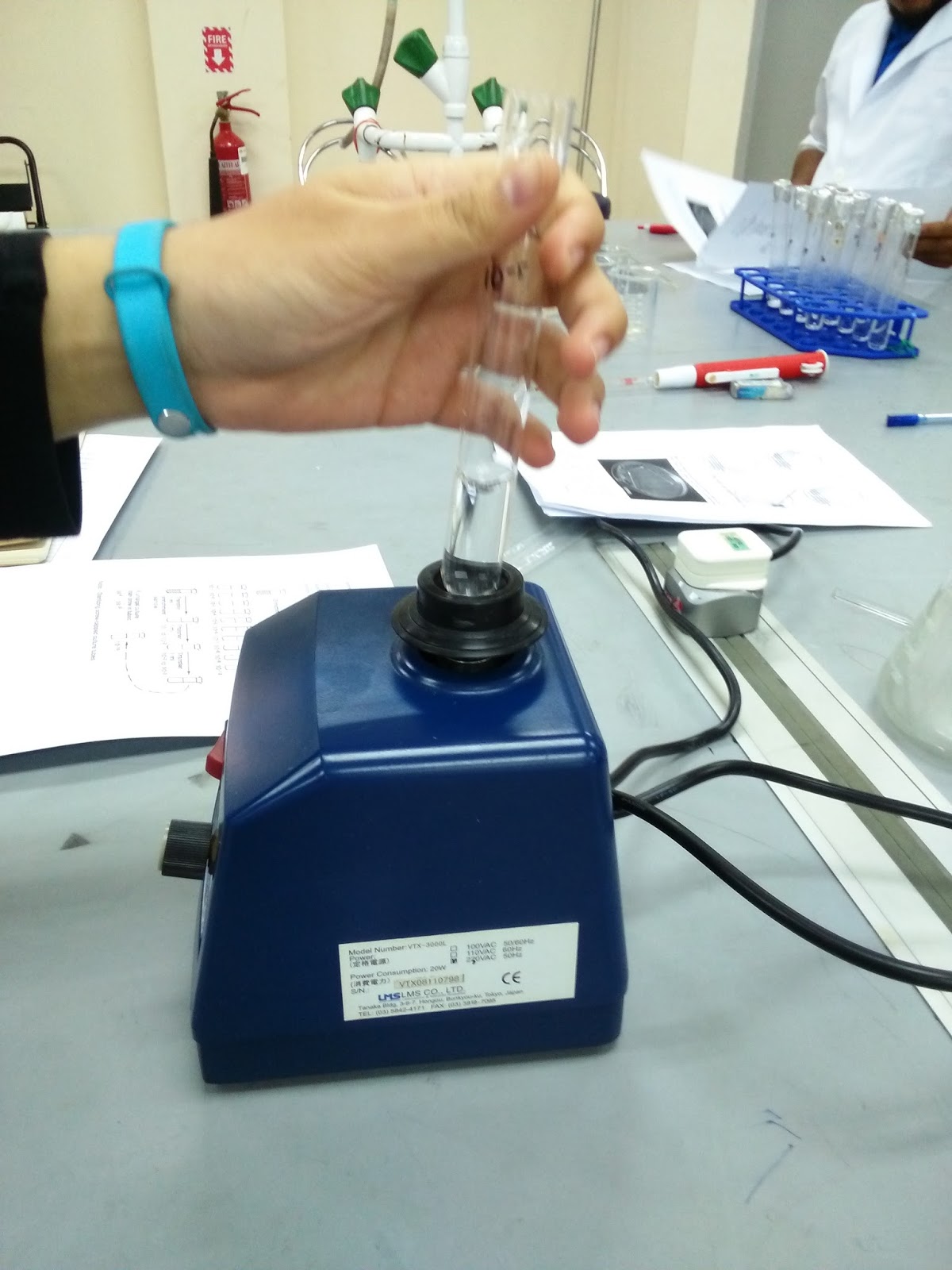
| Image 11: Centrifuge used to produce pallets of Culture X |
 |
| Image 12: Sterilising the wire loop before inoculating the pallet onto the agar plate. |
 |
| Image 13: an f/2 medium agar plate after agar streaking of Culture X. |
No comments:
Post a Comment